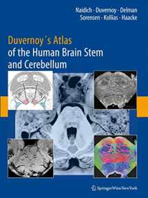
Duvernoy's Atlas of the Human Brain Stem and Cerebellum: High-Field MRI, Surface Anatomy, Internal Structure, Vascularization and 3 D Sectional Anatomy
Book details
Summary
Description
This atlas instills a solid knowledge of anatomy by correlating thin-section brain anatomy with corresponding clinical magnetic resonance images in axial, coronal, and sagittal planes. The authors correlate advanced neuromelanin imaging, susceptibility-weighted imaging, and diffusion tensor tractography with clinical 3 and 4 T MRI. Each brain stem region is then analyzed with 9.4 T MRI to show the anatomy of the medulla, pons, midbrain, and portions of the diencephalonin with an in-plane resolution comparable to myelin- and Nissl-stained light microscopy. The book’s carefully organized diagrams and images teach with a minimum of text.


We would LOVE it if you could help us and other readers by reviewing the book

