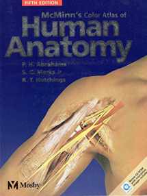
McMinn's Color Atlas of Human Anatomy: with STUDENT CONSULT Online Access
ISBN-13:
9780723432128
ISBN-10:
0723432120
Edition:
5
Author:
Ralph T. Hutchings, Peter H. Abrahams MBBS FRCS(ED) FRCR DO(Hon) FHEA, Stanley L Marks
Publication date:
2003
Publisher:
Mosby Ltd.
Format:
Paperback
392 pages
FREE US shipping
Book details
ISBN-13:
9780723432128
ISBN-10:
0723432120
Edition:
5
Author:
Ralph T. Hutchings, Peter H. Abrahams MBBS FRCS(ED) FRCR DO(Hon) FHEA, Stanley L Marks
Publication date:
2003
Publisher:
Mosby Ltd.
Format:
Paperback
392 pages
Summary
McMinn's Color Atlas of Human Anatomy: with STUDENT CONSULT Online Access (ISBN-13: 9780723432128 and ISBN-10: 0723432120), written by authors
Ralph T. Hutchings, Peter H. Abrahams MBBS FRCS(ED) FRCR DO(Hon) FHEA, Stanley L Marks, was published by Mosby Ltd. in 2003.
With an overall rating of 3.9 stars, it's a notable title among other
books. You can easily purchase or rent McMinn's Color Atlas of Human Anatomy: with STUDENT CONSULT Online Access (Paperback) from BooksRun,
along with many other new and used
books
and textbooks.
And, if you're looking to sell your copy, our current buyback offer is $0.32.
Description
This popular atlas maps out the structures of the human body and puts them in a clinical context. It incorporates an unrivalled collection of cadaveric, osteological, and clinical images with surface anatomy models, interpretive drawings, orientational diagrams, and diagnostic images. The 5th Edition features over 50 new dissection photos, many of which are taken from a distance to make them more recognizable in the lab setting. It also offers a more streamlined, user-friendly design, more clinical tips, and a companion CD-ROM with seven anatomical animations.
- Displays x-ray, MR, and CT images next to corresponding dissections, teaching readers to recognize anatomic structures in diagnostic images.
- Helps readers differentiate between similar structures on specimens (ie. veins, arteries, and nerves) with interpretive drawings.
- Offers orientational diagrams that make it easier to relate the dissections in the text to real experiences in the lab.
- Provides an unobstructed view of the artwork by using a numbered labeling system, which also allows readers to self-test by covering up the key and identifying each numbered structure.
- Includes over 50 new dissections photos, many of which are taken from a distance to make them more recognizable in the lab setting.
- Features a redesigned page layout that correlates illustrations, headings, and labels with more clarity.
- Uses more distinctly color-coded chapters to improve accessibility.
- Makes it easy to locate dissection sites on real life models with 50 new 'locator' images (surface anatomy models).
- Adds 20 new clinical tips (300 in all) that are referenced with icons on each page and then listed at the end of each chapter.
- Moves the Systemic Review to the front of the book, adding a series of anatomical cross-sections.
- Offers a PC and Mac compatible CD-ROM with seven anatomical animations (hand, shoulder, knee, foot, hip, spine, and head & neck) in QuickTime format. Allows users to add and remove layers of skin, muscle, etc.
- rotate animations for a complete view from all angles
- turn labels on and off to test recognition skills
- export images to their hard drive
- test themselves with multiple-choice questions linked to each label as well as with a separate quiz function
- link to www.fleshandbones.com. Icons in the book refer readers to the animations at appropriate points.


We would LOVE it if you could help us and other readers by reviewing the book
Book review

Congratulations! We have received your book review.
{user}
{createdAt}
by {truncated_author}


