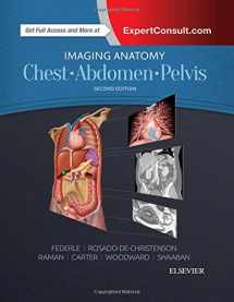
Imaging Anatomy: Chest, Abdomen, Pelvis
ISBN-13:
9780323477819
ISBN-10:
032347781X
Edition:
2
Author:
Siva P. Raman MD, Michael P. Federle MD FACR, Melissa L. Rosado-de-Christenson MD FACR, Brett W. Carter MD, Paula J. Woodward MD, Akram M. Shaaban MBBCh
Publication date:
2016
Publisher:
Elsevier
Format:
Hardcover
1192 pages
Category:
Anatomy
,
Basic Medical Sciences
FREE US shipping
Book details
ISBN-13:
9780323477819
ISBN-10:
032347781X
Edition:
2
Author:
Siva P. Raman MD, Michael P. Federle MD FACR, Melissa L. Rosado-de-Christenson MD FACR, Brett W. Carter MD, Paula J. Woodward MD, Akram M. Shaaban MBBCh
Publication date:
2016
Publisher:
Elsevier
Format:
Hardcover
1192 pages
Category:
Anatomy
,
Basic Medical Sciences
Summary
Imaging Anatomy: Chest, Abdomen, Pelvis (ISBN-13: 9780323477819 and ISBN-10: 032347781X), written by authors
Siva P. Raman MD, Michael P. Federle MD FACR, Melissa L. Rosado-de-Christenson MD FACR, Brett W. Carter MD, Paula J. Woodward MD, Akram M. Shaaban MBBCh, was published by Elsevier in 2016.
With an overall rating of 4.2 stars, it's a notable title among other
Anatomy
(Basic Medical Sciences) books. You can easily purchase or rent Imaging Anatomy: Chest, Abdomen, Pelvis (Hardcover) from BooksRun,
along with many other new and used
Anatomy
books
and textbooks.
And, if you're looking to sell your copy, our current buyback offer is $2.32.
Description
Designed to help you quickly learn or review normal anatomy and confirm variants, Imaging Anatomy: Chest, Abdomen, Pelvis provides detailed views of anatomic structures in successive imaging slices in each standard plane of imaging. Axial, coronal, sagittal, and 3D reconstructions accompany highly accurate and detailed medical drawings, assisting you in making an accurate diagnosis. Comprehensive coverage of the chest, abdomen, and pelvis, combined with an orderly, easy-to-follow structure, make this unique title unmatched in its field.
- Includes all relevant imaging modalities , 3D reconstructions, and highly accurate and detailed medical drawings that illustrate the fine points of the imaging anatomy
- Depicts common anatomic variants and covers common pathological processes as a part of its comprehensive coverage
- Provides a detailed overview of airway and interstitial network anatomy―the basis for understanding and diagnosing interstitial lung disease
- Features representative pathologic examples to highlight the effect of disease on human anatomy
- Includes plain radiography, the latest generation of multi-planar advanced cross-sectional MR and CT, ultrasound for pelvis/renal/liver/gallbladder, barium for GI tract, and much more
- Offers state of the art, detailed pelvic floor imaging and perianal/perirectal fistula imaging using high-resolution CT and MR, including 3T MR
- Expert Consult eBook version included with purchase. This enhanced eBook experience allows you to search all of the text, figures, images, and references from the book on a variety of devices.


We would LOVE it if you could help us and other readers by reviewing the book

{user}
{createdAt}
{truncated_title}
{author_block}
by {truncated_author}
