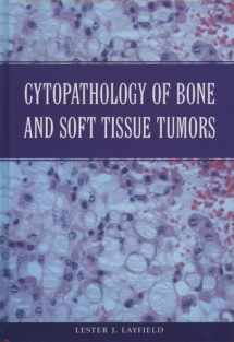
Cytopathology of Bone and Soft Tissue Tumors
ISBN-13:
9780195132366
ISBN-10:
019513236X
Edition:
1
Author:
Pamela Mason, Mehta, Parkin, Lester J. Layfield, Trobe, Parthenon, W. Holzgreve, D.A. Nyberg, Shankie, Bhugra, Mark Freed
Publication date:
2002
Publisher:
Oxford University Press
Format:
Spiral-bound
280 pages
Category:
Botany
,
Biological Sciences
FREE US shipping
Book details
ISBN-13:
9780195132366
ISBN-10:
019513236X
Edition:
1
Author:
Pamela Mason, Mehta, Parkin, Lester J. Layfield, Trobe, Parthenon, W. Holzgreve, D.A. Nyberg, Shankie, Bhugra, Mark Freed
Publication date:
2002
Publisher:
Oxford University Press
Format:
Spiral-bound
280 pages
Category:
Botany
,
Biological Sciences
Summary
Cytopathology of Bone and Soft Tissue Tumors (ISBN-13: 9780195132366 and ISBN-10: 019513236X), written by authors
Pamela Mason, Mehta, Parkin, Lester J. Layfield, Trobe, Parthenon, W. Holzgreve, D.A. Nyberg, Shankie, Bhugra, Mark Freed, was published by Oxford University Press in 2002.
With an overall rating of 4.1 stars, it's a notable title among other
Botany
(Biological Sciences) books. You can easily purchase or rent Cytopathology of Bone and Soft Tissue Tumors (Spiral-bound) from BooksRun,
along with many other new and used
Botany
books
and textbooks.
And, if you're looking to sell your copy, our current buyback offer is $0.49.
Description
Cytopathology of Bone and Soft Tissue Tumors provides the practicing pathologist with a single reference for describing and illustrating the cytologic features of musculoskeletal tumors. Using fine-needle aspiration (FNA), the approach of this work is encyclopedic: both relatively common and relatively rare lesions are depicted. It is expected that Cytopathology of Bone and Soft Tissue Tumors will serve to widen the usage of the FNA technique which can: substantially decrease patient morbidity; lessen the complications that arise due to other biopsy techniques; and shorten the time as well as expense required for diagnosis. The chapters on soft tissue lesions are organized by direction of differentiation. Each chapter documents both the common and uncommon lesions within the tissue group. Summaries of clinical findings are given along with the histopathologic and cytologic description. Key diagnostic points (in tabular form) and a discussion of the differential diagnosis complete each section. The section on skeletal lesions is organized along predominant cell type seen in smears. This approach facilitates grouping of lesions into diagnostically useful categories, allowing the pathologist faced with an unfamiliar lesion to rapidly access the portion of the text most useful for differential diagnosis. Within each chapter, the organization is similar to that within the soft tissue chapters. The introduction discusses technical concerns, limitations of the technique, and a diagnostic approach in both tabular and narrative form. Information on grading of soft tissue sarcomas completes the introduction.


We would LOVE it if you could help us and other readers by reviewing the book
Book review

Congratulations! We have received your book review.
{user}
{createdAt}
by {truncated_author}


