9780125476225-0125476221-Atlas of the Developing Mouse Brain: E17.5, P0 and P6

Atlas of the Developing Mouse Brain: E17.5, P0 and P6

ISBN-13: 9780125476225
ISBN-10: 0125476221
Author: Charles Watson, Hongquin Wang, George Paxinos AO (BA MA PhD DSc) NHMRC, Glenda Margaret Halliday, Yuri Koutcherov
Publication date: 2006
Publisher: Academic Press
Format: Spiral-bound 378 pages
FREE US shipping

Book details

ISBN-13: 9780125476225
ISBN-10: 0125476221
Author: Charles Watson, Hongquin Wang, George Paxinos AO (BA MA PhD DSc) NHMRC, Glenda Margaret Halliday, Yuri Koutcherov
Publication date: 2006
Publisher: Academic Press
Format: Spiral-bound 378 pages

Summary

Atlas of the Developing Mouse Brain: E17.5, P0 and P6 (ISBN-13: 9780125476225 and ISBN-10: 0125476221), written by authors Charles Watson, Hongquin Wang, George Paxinos AO (BA MA PhD DSc) NHMRC, Glenda Margaret Halliday, Yuri Koutcherov, was published by Academic Press in 2006. With an overall rating of 4.0 stars, it's a notable title among other Neuropsychology (Psychology & Counseling) books. You can easily purchase or rent Atlas of the Developing Mouse Brain: E17.5, P0 and P6 (Spiral-bound) from BooksRun, along with many other new and used Neuropsychology books and textbooks. And, if you're looking to sell your copy, our current buyback offer is $0.46.

Description

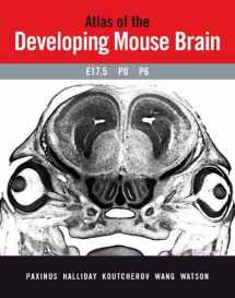
This atlas provides an accurate and detailed depiction of all brain structures at fetal stage E17.5, Day of birth, and Day 6 postnatal. In addition to brain structures, the atlas delineates peripheral nerves, ganglia, arteries, veins, muscles bones and other organs. It is an indispensable guide for the interpretation of nervous system changes in gene knockout and transgenic mice. The authors are recognized leaders in brain cartography, and the delineations and graphics link the atlas with other highly successful Paxinos atlases, complementing the catalog of brain atlases of highest quality available from Elsevier. There is currently no alternative of similar quality and extent on the market. · 43 photographs and drawings of Nissl-stained coronal sections of the brain of a fetal mouse at E17.5 days· 65 photographs and drawings of Nissl-stained coronal sections of the brain of a mouse on the day of birth· 73 photographs and drawings of Nissl-stained coronal sections of

Rate this book Rate this book

We would LOVE it if you could help us and other readers by reviewing the book