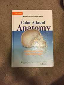
9781582558561-1582558566-Color Atlas of Anatomy: A Photographic Study of the Human Body

Color Atlas of Anatomy: A Photographic Study of the Human Body

ISBN-13: 9781582558561
ISBN-10: 1582558566
Edition: 7
Author: Johannes W. Rohen, Elke Lutjen-Drecoll, Chichiro Yokochi
Publication date: 2010
Publisher: Lippincott Williams & Wilkins
Format: Hardcover 532 pages
FREE US shipping
Rent
35 days
from $13.86 USD
FREE shipping on RENTAL RETURNS
Buy

From $35.40

Rent

From $13.86

Book details

ISBN-13: 9781582558561
ISBN-10: 1582558566
Edition: 7
Author: Johannes W. Rohen, Elke Lutjen-Drecoll, Chichiro Yokochi
Publication date: 2010
Publisher: Lippincott Williams & Wilkins
Format: Hardcover 532 pages

Summary

Color Atlas of Anatomy: A Photographic Study of the Human Body (ISBN-13: 9781582558561 and ISBN-10: 1582558566), written by authors Johannes W. Rohen, Elke Lutjen-Drecoll, Chichiro Yokochi, was published by Lippincott Williams & Wilkins in 2010. With an overall rating of 3.7 stars, it's a notable title among other Allied Health Professions (Anatomy, Basic Medical Sciences, Medical, Professional Tests) books. You can easily purchase or rent Color Atlas of Anatomy: A Photographic Study of the Human Body (Hardcover, Used) from BooksRun, along with many other new and used Allied Health Professions books and textbooks. And, if you're looking to sell your copy, our current buyback offer is $3.41.

Description

This Color Atlas of Anatomy features full-color photographs of actual cadaver dissections, with accompanying schematic drawings and diagnostic images. The photographs depict anatomic structures with a realism unmatched by illustrations in traditional atlases and show students specimens as they will appear in the dissection lab.

Chapters are organized by region in order of standard dissection, with structures presented both in a systemic manner, from deep to surface, and in a regional manner.

This edition has additional clinical imaging, including MRIs, CTs, and endoscopic techniques. New graphics include clinically relevant nerve and vessel varieties and antagonistic muscle functions. Many older images have been replaced with new, high-resolution images. Black-and-white dissection photographs have been replaced with color photography.

A companion website will include an Image Bank, interactive software (similar to an Interactive Atlas), and full text online.

Rate this book Rate this book

We would LOVE it if you could help us and other readers by reviewing the book